Please read at the bottom, Water and the Brain (the article includes two brief tutorial videos) to get an understanding of the brain and spinal column’s cerebral spinal fluid (CSF). But first, read the excerpts from the paper (below the photographs of artwork etc.) titled Robustness of the Mind-Body Interface: case studies of unconventional information flow in the multiscale living architecture. It provides example after example of extraordinary cases of typical and even exceptional mentally functioning individuals despite having missing considerable, nearly all in some cases, brain mass or some other severe neural disorder. It begins a bit scientific then soon quite layman comprehensible. As well initially, and disturbingly, the authors state: “Continued progress, in terms of positive impact on human flourishing and the ethics of relationships to unconventional beings (such as forthcoming enhanced humans, cyborgs, new brain-machine interface [transhumanism] require us to have a better understanding of the natural biological substrate of mind, and the space of the possible minds that can exist.”
We can’t ignore these frightening developments that require our attention in order to truly evolve intellectually, psychologically and spiritually without depending on or being merged with machines. The authors intent with the whole of the paper is to point out our natural highly complex, extraordinary evolving minds and physiology. I believe that (my own hypothesis) the information in the article suggests that water may very well be largely involved in central nervous system (thus entire biological) processing and conductivity (action potential in scientific parlance). In place of these areas of missing brain tissue is cerebral spinal fluid, water basically.
I’m a professional artist and I paint very realistically – it came naturally, was quite apparent when I was a child and I did not need training. Sometimes viewers find my work hard to distinguish whether my oil paintings are photographs or artistically rendered with paint, brush and canvas. There’s no computer technology involved. I look at photographs or an object before me and that is my source material from which I work. I told myself years ago that I must surpass what a photograph can do without rendering my artwork abstract or nonsensical. Now with AI artwork, the challenge is even greater. It is a challenge we alI face presently regardless of our professions and in many other ways our contributions to humanity.
detail from Manitou – Native American for the life force in all things. While painting this work and another, both spiritual in nature, I experienced spontaneous psychic metal bending. On a previous page on this website I included a digital book by physicist Sir Jagadish Chandra Bose (1858 – 1937) where he writes of findings of organic and inorganic (metal in particular) fatigue due to repeated and constant pressure. I thought perhaps this could be the cause of psychic metal bending. Uri Geller, the Israeli who initially brought attention to this unique phenomena, along with scientists studying his and others extraordinary mental faculties at Stanford Research Institute in the 1970s, will gently stroke a spoon or fork handle then it just seems to melt where he strokes it. My paint is gently applied to the canvas in strokes yet the metal was never touched by my hand nor did it come in contact with the canvas – just the brush hairs. And in the other painting, The Higher Realm Consultations the spontaneous metal bending of the palette knife rod holding the flimsy knife and wooden handle could not have been due to pressure while mixing oil colors on a slick wax paper surface. That part too, never touched the palette surface or was touched by my hand. Yet most importantly, and what I only recently factored in, is that in all the years I have been painting this phenomena never before happened until after I had learned of it and that it occurs, albeit rarely yet with others intentionally and as often as they wish, such as Uri Geller.
Can AI psychically bend metal?

.

.
******
Robustness of the Mind-Body Interface: case studies of unconventional information flow in the multiscale living architecture by Karina Kofman and and Michael Levin
Link to paper: https://osf.io/preprints/osf/fqm7r_v1
(you may have to wait for it to upload and though the article is 63 pages long (scrolling on the document not the slider on the right) over half of it is citations and references.
Excerpts from the paper:
“Treasure your exceptions! … Keep them always uncovered and in sight. Exceptions are like the rough brickwork of a growing building which tells that there is more to come and shows where the next construction is to be.”
– William Bateson
Page 8: How much neural substrate is required for normal function?
In humans, the brain is regarded as the basis of all mental attributes, arising from the function of neural architectures which were strongly shaped by evolutionary selection for the most adaptive functionality under constraints of energy demand and pregnancy risk for head size. Many aspects of consciousness and cognition are regarded as an emergent property of collective interaction of neuronal networks with a basis in the brain. It is widely accepted that many of the higher neurological functions, including somatosensory processing, motor processing, conscious experience, memory, and emotion, are all based in the brain and are the result of cerebral function.
Neuroscientists, such as Brodmann who pioneered brain mapping, have attributed functionality to particular brain regions, such as associating Broca’s area with speech production and associating the postcentral gyrus with the primary somatosensory cortex. In search of a seat of consciousness, modern neuroscience approaches attribute conscious experience and cognitive processing in large part to the Central Nervous System, especially in the cerebral cortex, where most of the thinking and processing is posited to take place. Conventional cognitive neuroscience assumes that consciousness, memory and cognition are generated by the brain and operate within the brain and CNS. This is strongly supported by many lines of argument, as seen in the event of traumatic brain injuries, neurodegenerative conditions, tumors, strokes, brain hemorrhage and the brain mass effect in contusions or focal lesions; these can dramatically impact cognition, intelligence, communication, and sensorimotor function and be life-threatening.
Page 9: However, there have been numerous clinical cases recorded in medical literature, in which patients have some kind of brain dysplasia (either congenital or acquired), reduced brain size, or some kind of massive lesioning, or a drastic absence of a brain matter, but exhibit normal cognitive development and function. These cases in human clinical literature demonstrate important challenges to our understanding of the relationship of functional cognition to a specific substrate.
Reduced brain mass or absent brain tissue unaccompanied by a loss of function
Here, we first dive into well-documented examples of patients with microcephalus, hydrocephalus, hemihydranencephaly, and hemispherectomy, or absence of cortical material, who are missing a large amount of brain matter but tend to lead ordinary lives with average, or above average, intelligence. Such cases demonstrate that brain size, amount of cortical tissue and neuron quantity, even within a single species, do not directly predict intelligence.
Page 10: The neurologist John Lorber identified numerous patients in his medical practice who had severe hydrocephalus as evidenced by their brain imaging results (around 1980). The ventriculomegaly went practically unnoticed and the patients led otherwise normal lives with very little brain tissue. For example, Lorber described a university honors mathematics student who had so much CSF that there was almost no white matter; the ventricular enlargement took up so much space and the actual brain was a thin mantle lining the inner skull. In contrast to the normal thickness of brain tissue between ventricles and outer cortex as being 4.5 cm, this student’s brain mantle measured a mere millimeter or so. Yet, this patient was socially normal, with an IQ of 126 and great academic achievement in math. Lorber’s patients had an utter lack of brain tissue which went totally unnoticed until imaging was taken.
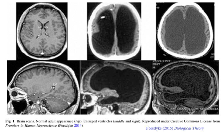
Above, the scans on the left is of a normal brain and the four scans on the right shows the cerebral cavities where cerebral spinal fluid, water mostly, is in place of where brain tissue ordinarily is.
Page 14: Dissociation of cognitive function from brain electrophysiology
Brain activity can be monitored via electrophysiological recordings, with the current paradigm driving the expectation that when electrical activity ceases, so does cognition. Next, we review cases suggesting mental activity despite documented, significant diminution or collapse of brain activity (with or without pathophysiology of the brain). This was, in some cases, paradoxically associated with periods of greater cognitive capacities, mental clarity, implicit learning, and vivid experiences.
Pages 17 & 18: Terminal Lucidity and Paradoxical Lucidity
Unlike salamanders and planaria, human brains are not thought to regenerate. The standard picture of advanced age and degenerative disease is that of consistent monotonic decline in cognitive capacities. However, this prediction is challenged by frequent reported cases of terminal lucidity, a phenomenon that has been reported in patients since the 18th century in many different retrospective case studies. Terminal lucidity occurs in patients with terminal conditions, including neurodegenerative conditions, minutes, hours, or days before death. Patients experience a sudden mental clarity, the return of memories and normal cognitive function shortly before dying, which seems impossible given the extent of their past state, and history of brain degeneration and damage. These coherent interactions can have a duration ranging from minutes to days, and most patients passaway within the next few days.
This phenomenon has been observed in the most enigmatic cases including Alzheimer’s patients, dementia patients, chronic schizophrenia patients, comatose patients, as well as those patients with brain abscess, brain tumors, strokes, and meningitis. For example, a case was reported of a young man with metastatic lung cancer, who was unable to speak or to move because the tumor had replaced his brain matter. However, in the hours before his death, he woke up and conversed with his family, said goodbye, then died shortly after.
******
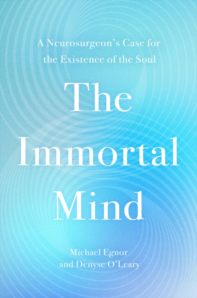
Another great source of similar cases is the book by neurosurgeon Michael Egnor and Denise O’Leary titled The Immortal Mind.
.
It includes the author’s own experiences, during surgery and with patients in his office and includes other cases similar to those referred to above. It’s quite readable and hard to put down. His position, different than mine regarding water’s role in cognition, yet neither excludes the other, is that consciousness, the immortal mind, is primary and despite severe neural damage, brain mass absence or deformities, this is why the individuals can and do in many cases, function normally. Dr. Egnor describes a case where he was operating on the brain of an awake patient (the brain has no feeling sensations) where he removed nearly all of her left pre frontal cerebral cortex and she was able to maintain a conversation with him the entire time. Neurosurgeons will do this in order to be very careful not to damage, to avoid, sensitive neural regions such as language facilitating areas – Broca’s area. However, no CFS likely was in or replaced the areas he removed while operating.
******
Water and the Brain
The percentage of water in brain tissue varies across different sources, but it is generally estimated to be around 65% to 80%. The rest of the human body is estimated to be roughly the same.
The human brain, the central nervous system (CNS), contains approximately one hundred billion neurons. There are other brain cells with glial cells being the most prominent. Glial cells support and maintain the function of neurons. There are several types of glial cells. For example oligodendrocytes, microglia, astrocytes and ependymal cells. Oligodendrocytes are responsible for forming the myelin sheath around axons which aids the transmission of electrical signals. Microglia act as an immune defense system in the brain, detecting and responding to damaged or unhealthy neurons. Astrocytes provide structural and metabolic support to neurons. Other types of glial cells are ependymal cells which form the membrane lining the four ventricles of the brain and the central column of the spinal cord. There are also satellite cells which are thought to provide neuron nutrient support and protection.
Cerebral Spinal Fluid (CSF) associated with autism spectrum Disorder (ASD) and Alzheimer’s disease.
Is it not possible that a diet with no sugar, very little salt and processed foods and drinking lots of pure clean water would flush the molecular toxins and metals (mercury, aluminum and thimerasal a crystalline mercurial antiseptic) in the cerebral spinal fluid and brain, not to mention the rest of the body (Americans are rapidly growing increasingly obese due to processed foods consumption) and heal Autism Spectrum Disorder, Alzheimer’s and ADHD – fasting in other words? I’m not suggesting this would be easy (more difficult for adults) but if I had an autistic child I would surely give it a try!
Science Direct 2025 article – Neuroscience and Biobehavior Reviews
Abstract: Autism Spectrum Disorder (ASD) is a range of neurodevelopmental conditions characterized by impaired social interaction, learning, and restricted or repetitive behaviors. The underlying causes of ASD are still debated, but researchers have found many physiological traits like gut problems and impaired immune system to help understand the etiology of ASD. Cerebrospinal fluid (CSF) plays a critical role in maintaining the homeostasis of the neuronal environment and has, therefore, been analyzed in multiple conditions impacting the central nervous system. The study of CSF is crucial to understanding neurological disorders as its composition changes with the disorders, and these changes may indicate various disorder-related physiological mechanisms. For this systematic review, we searched PubMed, Scopus, and Web of Science for studies published between 1977 and 2025 and selected 49 studies after manual screening. ”
The bottom third of the article is filled with links to numerous reports researching possible links between autism and the cerebral spinal fluid.
Source: Science Direct article: Cerebrospinal Fluid Findings of 36 Adult Patients with Autism Spectrum Disorder
https://www.sciencedirect.com/science/article/pii/S0149763425002027
(1)
Autism in Children
Another article about autism in small children features a brain scan showing an abnormally greater amount of CSF in a young child. The image below is of a 6 month old infant who at 2 years was diagnosed as autistic. Children typically exhibit autism syndrome disorder (ASD) at 2 to 4 years of age. The article suggests that ASD is caused by a prenatal birth defect of the brain due to the CSF volume abnormality. There are numerous scientific reports online adamantly denying that vaccines are at all responsible for the current and tragic epidemic of autism in children – now 1 in 33 children, mostly boys, whereas prior to the mid 1980s it was 1 in 10,000. Initially it was claimed that the increase was due to misdiagnosis, not properly diagnosing autism for decades, but that claim has since been found to be untrue.
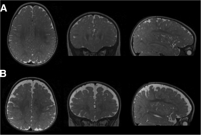
The brain scans in the top row are of a normal 6 month infant’s cranium and the bottom scans are of a 6 month old infant also at 6 months with a high volume of CSF who at two years of age was diagnosed with ASD.
Autism in Adults
“In total, more of the ASD patients (44%) presented abnormal findings in CSF basic diagnostics compared to controls (18%; p = 0.013). A subgroup of the patients with adult ASD showed indication of a blood–brain barrier dysfunction, and two patients displayed an intrathecal (delivered to the spinal canal) synthesis of anti-GAD65 antibodies [proteins produced by the immune system associated with various neurological syndromes]; thus, the role of these antibodies in patients with ASD should be further investigated.“
Source: 2020 NIH PubMed report – Cerebrospinal Fluid Findings of 36 Adult Patients with Autism Spectrum Disorder
https://pmc.ncbi.nlm.nih.gov/articles/PMC7349103
(2)
Diet and Cerebral Spinal Fluid
Diet can significantly affect cerebrospinal fluid (CSF) amino acid (molecular components of proteins) levels and other biomarkers observed in individuals with normal cognition (NC) and those with mild cognitive impairment (MCI). A study found that a high saturated fat/glycemic index (HIGH) diet led to increases in CSF amino acids valine and isoleucine, while decreasing arginine, compared to a low saturated fat/glycemic index (LOW) diet. These changes were more pronounced in individuals with MCI than in those with NC. Additionally, the study noted that changes in CSF amino acids were correlated with changes in Alzheimer’s disease biomarkers, indicators, such as Aβ42, total tau, and p-Tau181, with distinct patterns observed based on diet intervention and cognitive status.
Aβ42, Tau, p-Tau181 are biomarkers used in the diagnosis of neurodegenerative diseases, particularly Alzheimer’s disease (AD).
Aβ42 refers to the amyloid-beta 42 peptide, [peptides are smaller and less complex chains of amino acids than proteins] which is a key component of the extracellular neuritic plaques associated with AD pathology. Total tau (t-tau) is a protein that is present in the cerebrospinal fluid (CSF) and reflects the extent of neuronal death. p-Tau181, or phosphorylated tau at threonine 181, is a form of tau that is more closely associated with neuro-fibrillary tangle formation in AD. These biomarkers are often measured in CSF to help in the diagnosis and diagnosis of AD from other neurodegenerative disorders.
Another study highlighted that dietary interventions can influence CSF markers of Alzheimer’s disease. For instance, a HIGH diet was associated with increases in insulin concentration and lipid parameters, while a LOW diet had the opposite effect. The study also found that the HIGH diet induced changes in CSF Aβ42 concentrations that were consistent with amplified AD pathophysiologic changes, whereas the LOW diet had a suppressive effect.
These findings suggest that diet plays a crucial role in modulating the CSF composition and may have implications for the prevention and management of neurodegenerative diseases like Alzheimer’s. The specific effects of diet on CSF can vary depending on the individual’s cognitive status and the type of diet consumed.
(3)
Medical News Today article: Your brain health may benefit more from cycling than walking, study finds.
At the study’s conclusion, researchers found that participants who regularly rode a bicycle for transportation had a lower dementia risk compared to participants who walked, drove, or took the bus.
“Our key finding: Participants using cycling/mixed-cycling modes had a 19% lower all-cause dementia risk and 22% lower Alzheimer’s disease risk vs. nonactive modes,” Chen explained. “This likely stems from cycling’s higher aerobic intensity and cognitive engagement (e.g., navigation, coordination), which may enhance neuroplasticity more than walking alone.”
Additionally, Chen and his team discovered that participants who regularly rode a bicycle for transportation, or mixed in bicycle riding with other modes of transport, were able to retain more volume in the hippocampus of the brain. “Cycling/mixed-cycling was significantly associated with greater hippocampal volume. This matters because the hippocampus is central to memory formation and is among the first regions damaged in dementia. Retaining its structure may delay cognitive decline.”
— Liangkai Chen, PhD
(4)
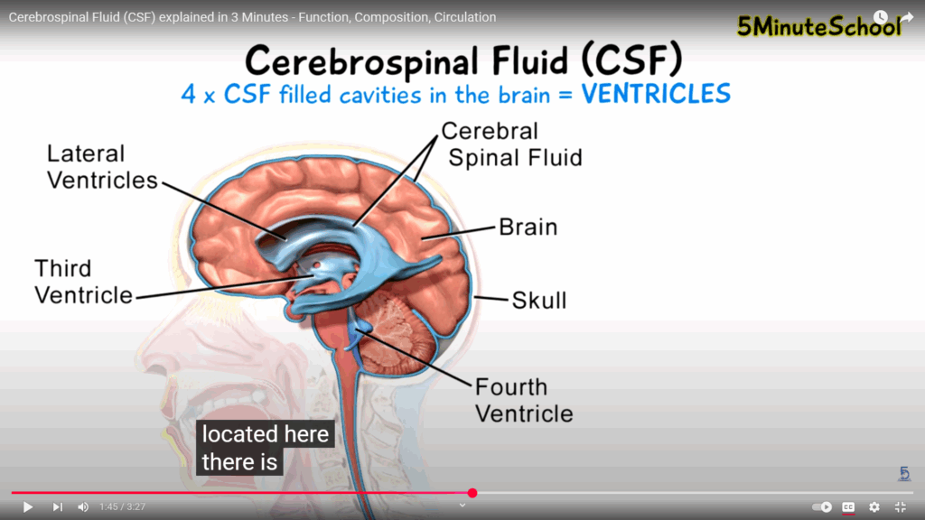
Brief tutorial by 5 Minute School: Cerebrospinal Fluid (CSF) explained in 3 Minutes – Function, Composition, Circulation:
Notes from the video – quite helpful to read prior to watching. Also some additional information:
The cerebral-spinal fluid is a liquid that protects the brain from physical and chemical damage. It contains substances that are both organic and inorganic. It circulates through the four ventricles of the brain, subarachnoid space and central canal of the spinal cord.
The total volume of CSF in a healthy adult is 80 –150 mL and production of the CSF is approximately 550mL per day and recycles approximately 3-4 times per day. 4 X 150 = 600mL – a bit high according to the figures presented in second more detailed, yet very good, video [link below]. Also mentioned in the second video is that the CSF also has homeostatic and lymphatic properties (protects against disease). The second video is far easier to follow (especially if you turn on the CC function) if the first is watched.
Contents of cerebrospinal fluid: The CSF is composed of water, sugars, proteins, lipids, and electrolytes, and it has a lot of the same components as plasma, which is the liquid part of your blood. However, CSF contains much lower amounts of protein than plasma does. Additionally, CSF consists predominantly of 99% water, and 1% accounts for electrolytes, proteins, neurotransmitters, and glucose.
Electrolytes defined: An electrolyte is a substance that conducts electricity through the movement of ions, but not through the movement of electrons. This includes most soluble salts, acids, and bases, dissolved in a polar solvent like water. Electrolytes are substances that have a natural positive or negative electrical charge when dissolved in water. They are nonmetallic electric conductors in which current is carried by the movement of ions. Electrolytes are minerals in your blood and other body fluids that carry an electric charge.
(5)
1) CSF provides a suitable chemical environment for neural signaling. If the chemical substances, the positive and negatively charged ions that facilitate action potentials and postsynaptic potentials, are out of balance this can seriously disrupt these nerve potentials.
2) CSF provides physical protection for the brain and spinal column in the cranial cavity and vertebral canal as a shock absorbing medium.
3) CSF is involved in the exchange of nutrients and waste products between the nerve tissue and blood.
4) CSF is produced in the choroid plexus (the CPE) shown here below as pale yellow (box-like) cells on the left which form the blood capillary transport networks in the walls of the ventricles. The ependymal cells (shown on the right are a specialized type of neural tissue found lining the central canal of the spinal cord and cerebral ventricles. The small encircled parts with orange dots are vesicles filled with CSF substances from the blood plasma (separately depicted red blood cells are on the left) being trafficked into the CSF ventricles and are what form the CSF. Brain parenchyma is brain tissue (shown here is an oligodendrocyte, astrocytes and neurons – there are others).
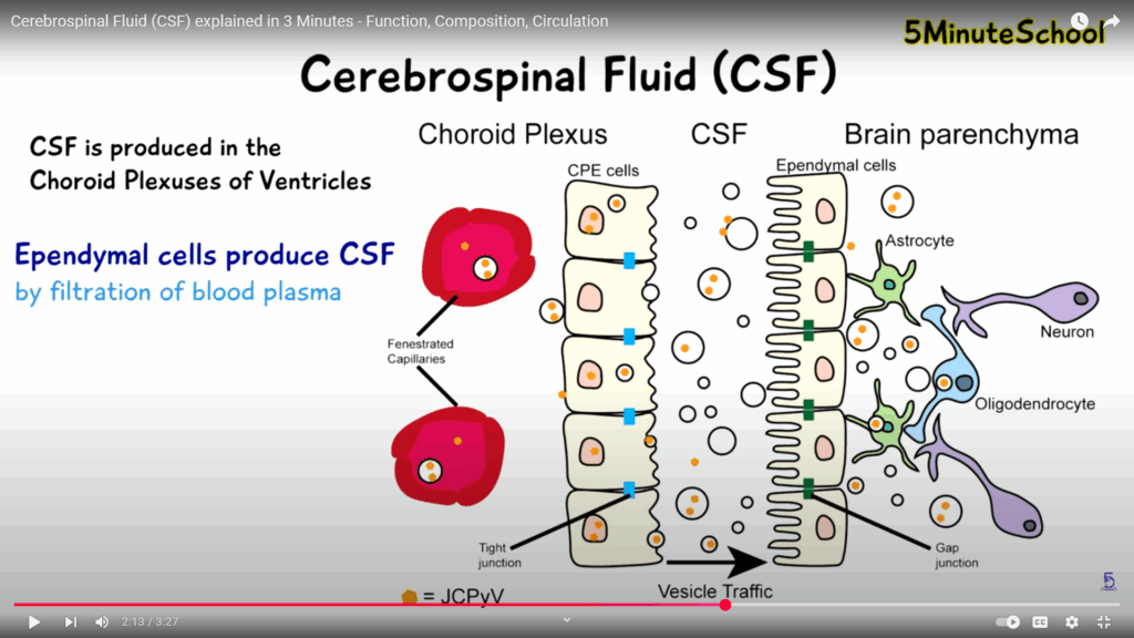
5) The small blue boxes of the CPE (choroid plexus) represents the tight junctions between the CPE cells which forms the blood-brain barrier allowing only certain substances to pass into the CSF. This is to protect the brain from toxic contaminants.
6) The CSF flows through the ventricles into the subarachnoid space (light blue layer) by the median and lateral apertures (lower left and right above the fourth ventricle labeled below) and some through the central canal of the spinal cord.
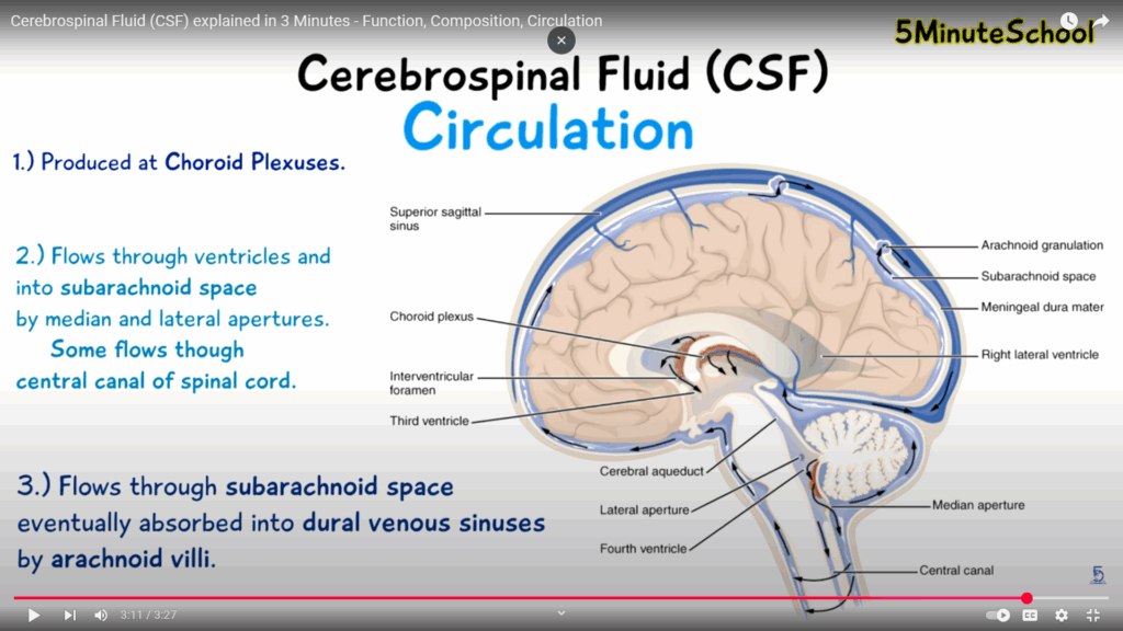
7) CSF Flows from the subarachnoid space (light blue layer) into the dura venal sinuses (the superior sagittal sinus shown on the upper left) by the arachnoid villi (see two light blue arachnoid granulations with arrows on upper right) flowing through the meningeal dura matter (the in between light gray layer).
8) CSF also assists in the removal of brain metabolism waste products, such as peroxidation products, glycosylated proteins, excess neurotransmitters, debris from the lining of the ventricles, bacteria, viruses and otherwise unnecessary molecules. Accumulation of such unnecessary molecules, seen in aging and some neuro-degenerative diseases, interferes with neuronal functioning of the brain. The disruption of cerebral physiology experienced with the disruption of the hydrodynamics or composition of CSF suggests the importance of CSF functioning.
For a more detailed CSF tutorial video titled: Cerebrospinal fluid physiology mbbs 1st year | CSF circulation and functions by a Sri Lanka professor which is easier to follow after viewing and reading the notes from the first brief video. It also helps to use the CC function.
https://www.youtube.com/watch?v=XnL8p7c_x1M
I can be contacted at: leslietaylorpaintings@gmail.com